


EQUIPMENTS
State of The Art Equipments
ALCON CENTURION VISION SYSTEM AND AMO PHACOEMULSIFICATION
Centurion offers ‘Active Fluids’ which monitores the eye pressure real time and thus gives the eye to nearest to normal conditions during every step of surgery – Safer surgery Specially designed for smoother cutting and excellent efficiency with the advent of blended phacoemulsification combining simultaneous transverse and longitudinal needle movement during cataract surgery – Lesser manipulation Gives excellent postoperative vision by using minimal ultrasonic power during surgery – Faster recovery
GREEN LASER PHOTOCOAGULATOR
Helps in advanced diabetic eye care. Prevents complications like bleeding in the eye and detachment of retina. Painless procedure – involves a light to help in treatment
OPTICAL COHERENCE TOMOGRAPHY SYSTEM (O.C.T.)
This is like a C.T. Scan of the eye without radiation or injection of dye. Retina and cornea can be evaluated layer by layer. Extremely useful for LASIK patients, before and after operation. Necessary before and after complicated operations for the anterior segment of the eye. Helps in monitoring n diabetic patients, Age related retinal conditions and other blood vessel related diseases.
ZEISS IOL MASTER
Gold standard all over the world for cataract surgery evaluation. Extremely useful and advanced technique required for premium IOLs and routine cataract surgery patients. Patient satisfaction increases due to independence from glasses.
TOPCON FUNDUS CAMERA WITH ANGIOGRAPHY
All retinal conditions can be photographed, processed and documented. Serial photographs over a period of time, helps in monitoring and treatment modification. Fundus Fluorescein angiography is an indispensable tool in diagnosis and management of retinal conditions.
OCULUS CORNEAL TOPOGRAPHY
Useful in selecting and fitting of contact lenses, routine and custom made lenses. Specifically required for LASIK operations . Advanced tool to diagnose and treat corneal diseases like keratoconus Before corneal operations like transplantation
HUMPHREY PERIMETRY
Helps to systematically measure the differential light sensitivity in the visual field function. Used to diagnose and monitor glaucoma and neurological defects. It measures peripheral field of vision which is lost first in glaucoma. Also useful in diagnosing and managing of tumors of brain, infarcts and bleeds.
YAG LASER
Helps to restore vision after posterior capsular opacification. Treatment of choice for angle closure glaucoma. The procedure is painless.
AUTO-REFRACTOR WITH KERATOMETER
Gives the corneal curvature and refractive power of the eye. Used for spectacle dispensing and contact lenses. Needed for preoperative cataract and LASIK work up when we want to correct preoperative astigmatism.
NONCONTACT TONOMETER WITH PACHYMETER
Gives the Intra ocular pressure(IOP) measurement and thickness of the cornea. Useful for diagnosis and management of glaucoma. Can be used even in chilDr.en, anxious and uncooperative patients.
PERKINS APPLANATION TONOMETER AND PASCAL DYNAMIC CONTOUR TONOMETER Applanation tonometery is the gold standard world wide for measuring eye pressure Dynamic contour tonometer gives accurate readings even in thinner or thicker corneas. Especially after corneal surgeries or in keratoconus VACCUM AUTOCLAVE AND ETO STERILISER It is the latest device used to sterilize the equipments by subjecting them to high pressure saturated steam at 1210c. Helps to maintain 100% sterilization in intraocular surgery ETO is a useful and safe method of instrument sterilization used all over the world. A-B SCAN USG Tried and tested method in IOL power calculation B-scan Helpful in diagnosis of foreign bodies inside eye, retinal detachments and bleeds. AUTO-LENSOMETER It is used to ascertain the spectacle power- Sovereign Classic Phaco Machine
- Associate 2500 Phaco with Vitrectomy
- Zeiss and Moller Wedel Microscopes
- GYC 1000 Green Laser for Photocoagulation by Nidek,
- Fundus camera and imaging by Topcon
- Optical Coherence Tomography by Optovue
- BIOM by Oculus,
- IOL Master by Zeiss,
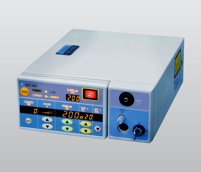
Green Laser Photocoagulator Gyc 1000 From Nidek, Japan
The art of Eye Care.
Efficient, safe Photocoagulation by 532 nm Laser beam. Frequency double diode pumped solid state laser. Laser is absorbed by pigment epithelium and Haemoglobin. Low absorption by xanthophyll pigment at macula. Exposure time adjustable in 0.05 second increments.
Helps in advanced Diabetic eye care.
Prevents complications like Bleeding in the eye and detachment of Retina.
Timely management by laser maintains the vision in diabetic patients and prevents them from becoming blind.
This is also required in operations of Retina.
Regular follow up and laser treatment avoid the necessity of operation for Retinal complications.
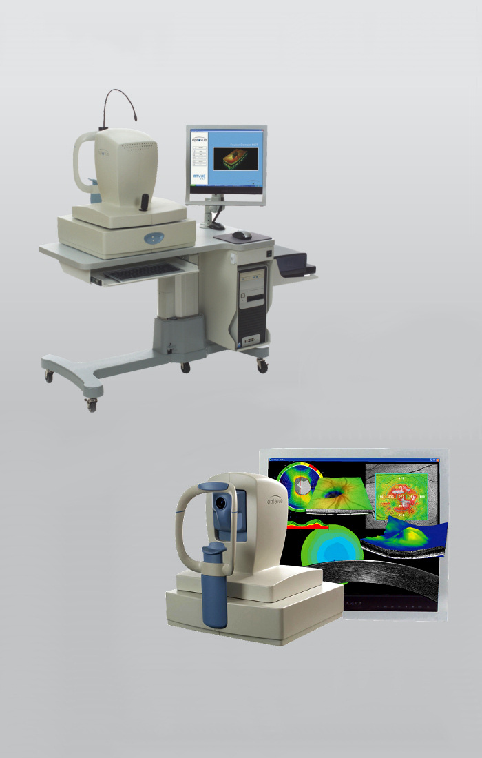
RT Vue Fourier –Domain Optical Coherence Tomography System (O.C.T.) by Optovue USA.
RETINA:
High Speed, high resolution of 5 microns for discrete layers of Retina. Earlier detection of Retinal Pathology allows earlier intervention and better outcomes for patients. Infra-Red fundus image is obtained without flash or visible light. This is like a C.T. Scan of the eye without radiation or injection of dye. Being objective test, prevents patient dependent mistakes and variations.
For early glaucoma detection and management tool. Optic Nerve head mapping scan shows real progression and accurate, repeatable analysis. Ganglion Cell complex (GCC) normative analysis detects significant changes associated with glaucoma with high sensitivity. 3 D Optic scan gives a overview and precise disc changes. CORNEA:
High Resolution imaging of cornea for documentation and precise measurements of anterior segment of the eye, Corneal thickness measurement (Pachymetry ), anterior Chamber angle measurement and epithelium / Lasik flap measurement can be done. Keratoconus analysis, Phakic IOL vault measurement Shunt / Implant imaging is possible. Extremely useful for LASIK patients, before and after operation. Necessary before and after complicated operations for the anterior segment of the eye.
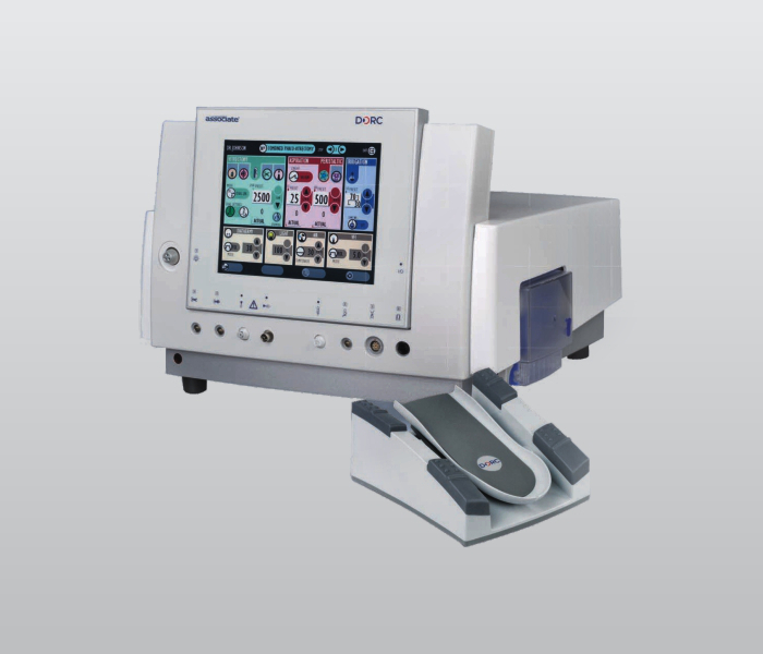
ASSOCIATE 2500 BY DORC (Netherlands)
Anterior and Posterior Segment Phaco with Vitrectomy with Dual Pump (Peristaltic and Ventury) Flow and Vacuum control. Dual linear 3D high speed Vitrectomy. Softsonic Phaco with 1.8mm micro incision technology.
Useful for all operations of Retina like detachment or complications due to Diabetes. Required after post operative complications of cataract surgery to save the eye and maintain good vision. Necessary to treat any complications for routine eye surgeries, to give excellent post operative result.
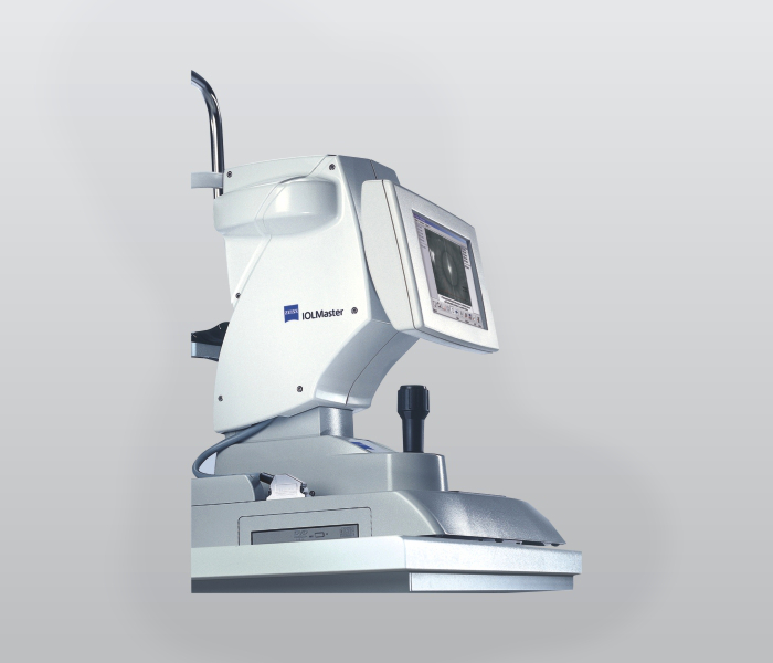
IOL MASTER BY ZEISS, GERMANY
For selecting the Right IOL to meet individual patient's needs, IOL Master is the benchmark for precision biometry. Accurate measurements of axial length and Keratometry with built in IOL power calculation options for latest formulas including premium IOL's and post LASIK Cases.
Extremely useful and advanced technique required for premium IOLs and routine cataract surgery patients.
Patient satisfaction due to spectacle non dependence increases.Patient can really enjoy the spectacle freedom due to multifical, Toric and variety of other IOLs because of accuracy achieved by the help of this machine.
Gold standard all over the world for cataract surgery evaluation.
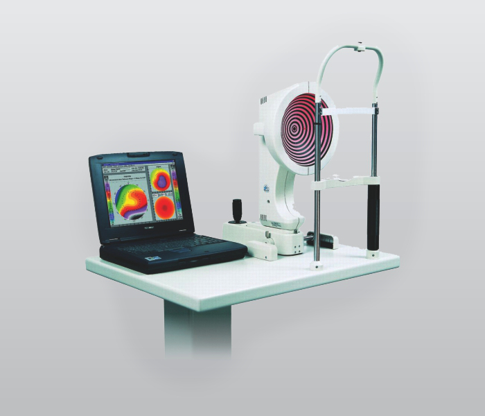
CORNEAL TOPGRAPHY BY OCULUS
Combination of Keratometric and corneal topography measurement. High resolution of cornea by measuring 22000 points. Digital image transmission and non contact measurement . Also used for Keratoconus detection and Contact Lens Fitting Pupillometry for measurement of Pupil in light and dark conditions. Capable of analysis and comparisons of all screenings.
Useful in selecting and fitting of contact lenses, routine and custom made lenses.
Specifically required for LASIK operations to reduce spectacle number, before and after surgery.
Advanced tool to diagnose and treat corneal diseases and before corneal operations including eye donation transplantation.
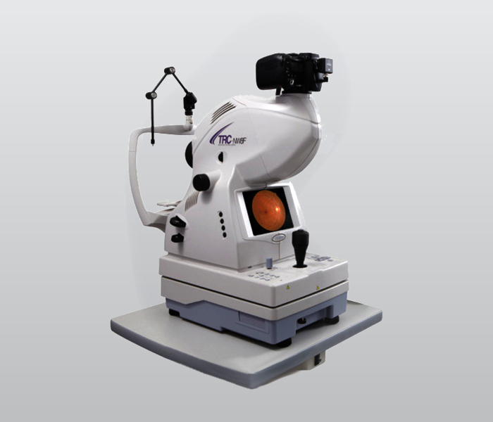
FUNDUS CAMERA BY TOPCON, JAPAN (TRC-NW8F MyDr.iatic / Non MyDr.iatic)
High Resolution fundus imaging system for screening of Diabetic Retinopathy and other retinal disorders like. Glaucoma, Age related macular degeneration, Macular holes etc. Auto focus, auto capture for colour and monochrome images of retina and anterior segment of the eye. 12.3 megapixel camera provides 45D field of view. Fluorescein angiography images are processed by IMAGE net 4 software System.
All diseases and problems of Retina can be photographed, processed and documented.
Serial photographs over a period of time, shows improvement or deterioration and helps in modifying the treatment.
Requirement of laser therapy can be decided by the angiography reports.
Most useful in patients of Diabetes with Retinopathy.
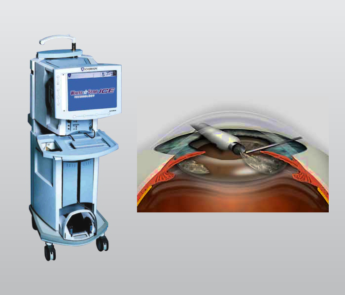
WHITE STAR SOVEREIGN CLASSIC PHACOEMULSIFICATION MACHINE BY A.M.O., U.S.A
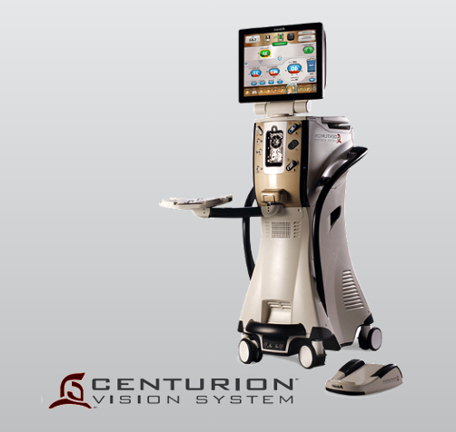
CENTURION VISION SYSTEM